

Chiari Malformations Neurosurgery Geeky Medics
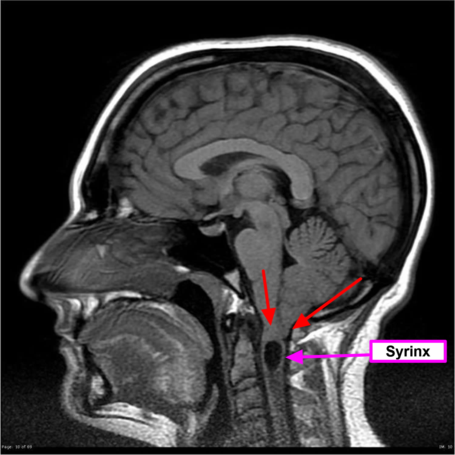
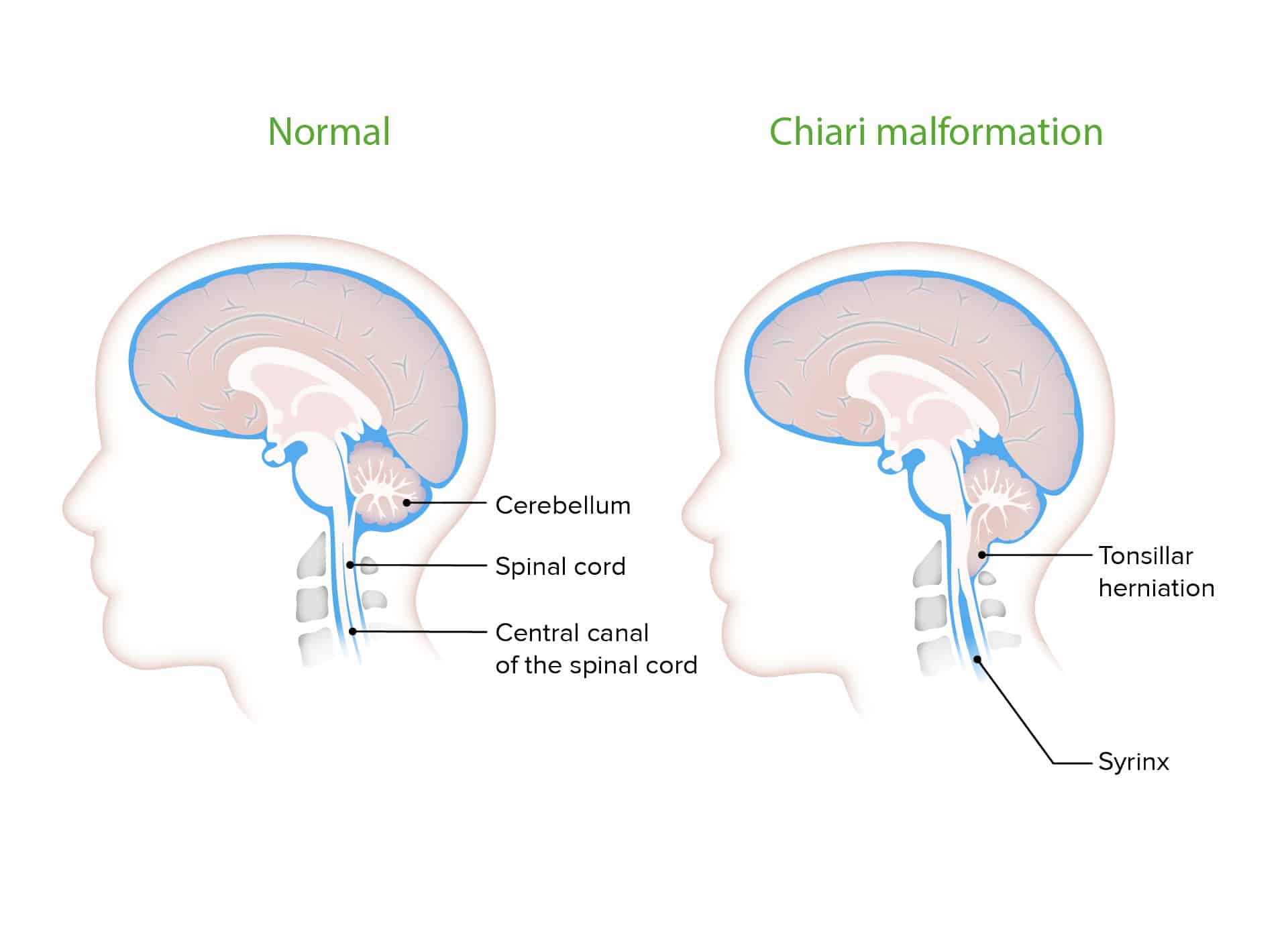
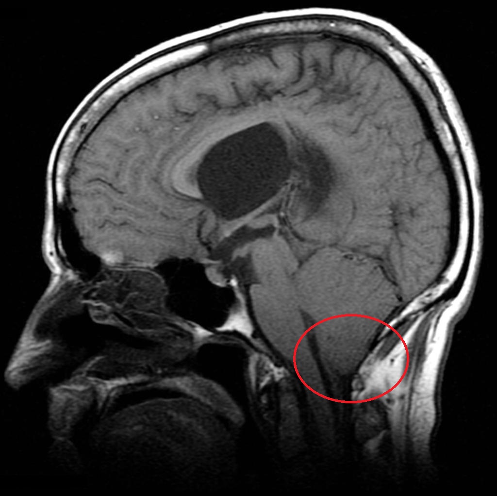
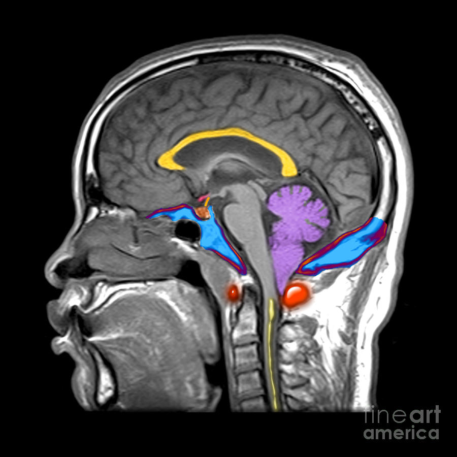
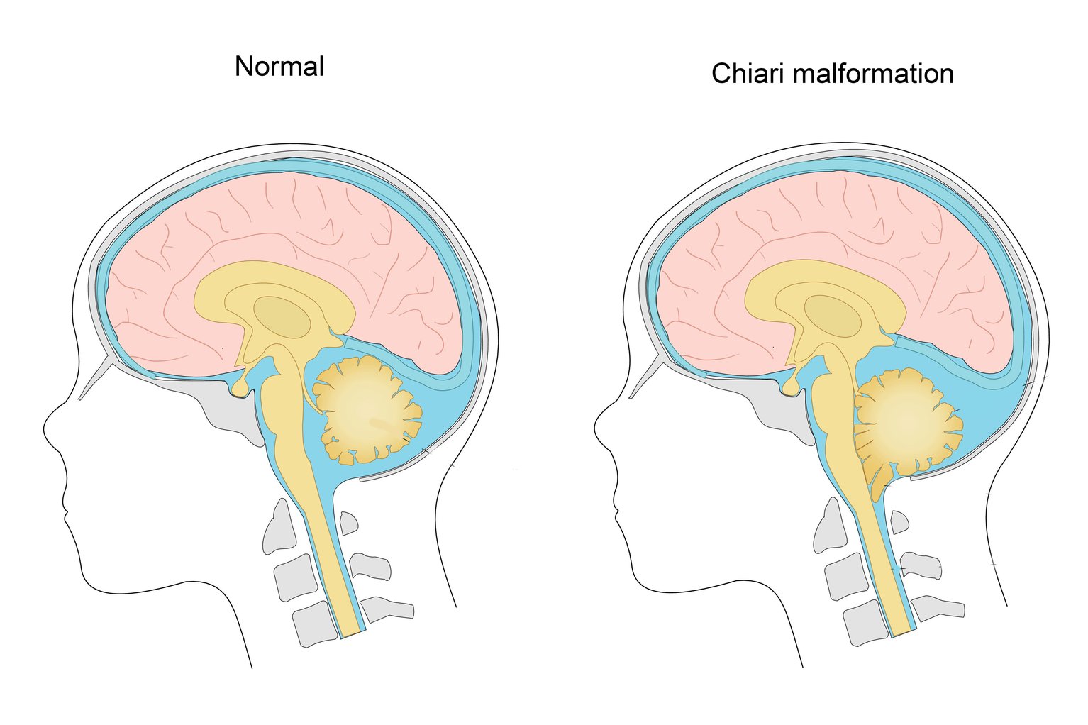
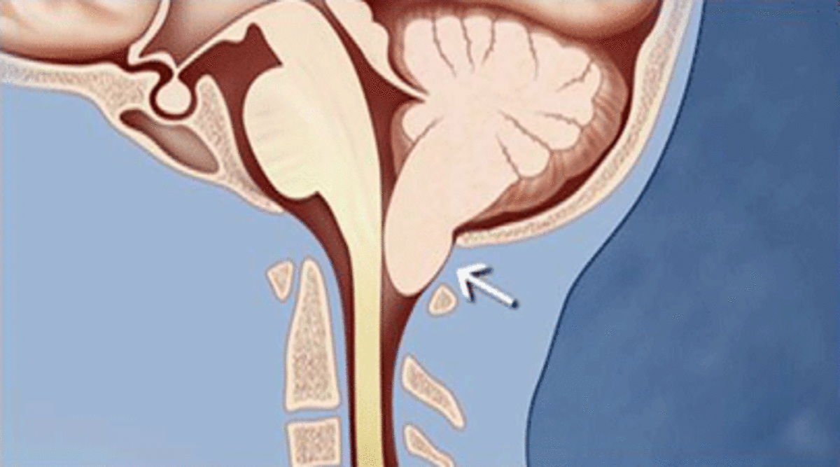
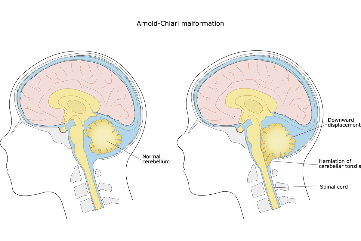
Chiari malformation type I (CM-I) is a congenital hind-brain defect characterized by herniation of the cerebellar tonsil through the foramen magnum with subsequent impairment of
Mal Formação De Chiari ENSINO
Causes. Chiari malformation type 1 occurs when part of the skull is too small or is misshapen. This part of the skull contains the area of the brain called the cerebellum. The skull puts pressure on and crowds the brain. As a result, the lower part of the cerebellum known as the tonsils are pushed into the upper spinal canal.

Malformation de Chiari
Clinical findings and magnetic resonance (MR) images in 68 patients with Chiari I malformations were retrospectively analyzed to identify those radiologic features that correlated best with clinical symptoms. A statistically significant (P = .03) female predominance of the malformation was observed, with a female: male ratio of approximately 3:2. Associated skeletal anomalies were seen in 24%.

Craniocervical for chiari malformation Resource Library Sheffield Children's
Fiche de cours collaborative au sujet de la maladie Malformation de Chiari éditée par le site Radeos Espace réservé aux professionnels de santé En application de la loi du 29 décembre 2011 relative au renforcement de la sécurité sanitaire et des produits de santé (dite loi « Bertrand »), je certifie être un professionnel de santé et être ainsi autorisé à accéder à cet espace.

Chiari malformation treatment
A Chiari malformation, previously called an Arnold-Chiari malformation, is where the lower part of the brain pushes down into the spinal canal. There are 4 main types, but type 1, called Chiari I, is the most common. In someone with Chiari I, the lowest part of the back of the brain extends into the spinal canal.
Chiari Malformation Hart Garner, MD
Le centre de référence « Chiari et malformations vertébrales et médullaires » rares (C-MAVEM) regroupe plusieurs équipes françaises, avec notamment différents services de neurochirurgie, d'urologie et un centre anti-douleur.Elles sont toutes impliquées dans le traitement des maladies rares de la moelle épinière dont les malformations de Chiari et les syringomyélies, de l.
Chiari Malformations Neurosurgery Geeky Medics
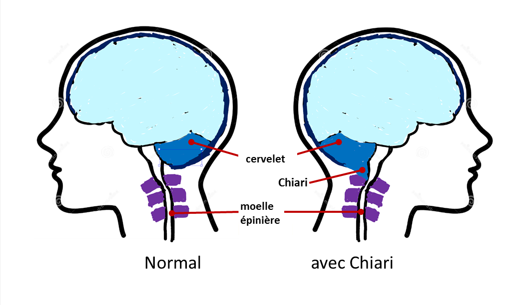
La malformation de Chiari est une malformation rare congénitale de la partie postèrieure du crâne.Le cervelet - qui est situé à l'arrière de la tête - est responsable de l'équilibre et de la coordination des mouvements. Les tonsilles (aussi appelées amygdales) cérébelleuses sont inhabituellement basses et s'engagent au travers du trou occipital (aussi appelé foramen magnum).
Chiari I Malformation MRI Photograph by Living Art Enterprises and Photo Researchers Pixels Merch
Chiari malformations are a heterogeneous group of hindbrain anomalies. Six different malformations are described. Most common are Chiari 1 malformation (CM1) and Chiari 2 malformation (CM2, also termed "Arnold-Chiari malformation") and are the focus of this review. These are rare conditions, but symptoms may impair quality of life in both.
Chiari malformation NHS
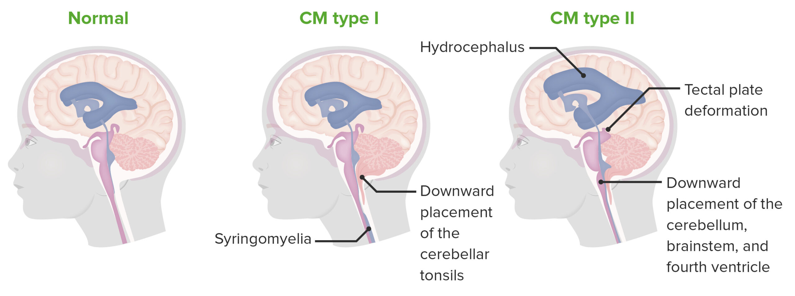
associée. La malformation de Chiari type II est une descente tonsillaire secondaire à un dysraphisme (spina bifida), en général une myéloméningocèle. D'autres types de malformations de Chiari ont été décrits (types 0, 0.5, 1.5, etc.), sans que cela ne fasse l'objet d'un consensus parmi les spécialistes.

Mal Formação De Chiari ENSINO
La malformation d'Arnold-Chiari est une anomalie congénitale qui affecte la partie inférieure du cerveau, appelée le cervelet. Dans cette condition, une partie du cervelet descend dans le canal rachidien, un espace normalement réservé à la moelle épinière. Cette descente peut entraîner une compression du cerveau et de la moelle.
Caring For A Child With Chronic Illness, Chiari Malformation WeHaveKids
Malformation de Chiari. Ce protocole national de diagnostic et de soins (PNDS) explicite aux professionnels concernés la prise en charge diagnostique et thérapeutique optimale et le parcours de soins d'un patient atteint de malformation de Chiari. Il a été élaboré par Centre de référence C-MAVEM Chiari, syringomyélie et malformations.
Malformación de Chiari Neurocirujano especialista columna
1. Introduction. Chiari malformations (CMs) comprise a series of neurodevelopmental disorders characterized by a descent of the cerebellar tonsils through the foramen magnum (FM) [1,2,3,4,5].CM type 1 (CM1) was first described by the Austrian pathologist Hans Chiari in two papers published in 1891 and 1895 [2,3,4].Nevertheless, the term "Arnold-Chiari malformation" must still be introduced.

Chiari Malformation — Matthew Mian, MD
At the end of the nineteenth century, two pathologists, Julius Arnold (1835-1915) and Hans Chiari (1851-1916), described a complex clinical and pathological condition involving deformity of the cerebellum and brainstem, in children. Chiari malformations are now defined as a spectrum of hindbrain abnormalities involving the cerebellum, brainstem, skull base, and cervical cord. According to the.
Chiari Malformations Types & Treatments Michigan Head & Spine Institute Blog
Chiari malformation (CM), one of the congenital disorder in Central nervous system (CNS) anomalies, is characterized by downward displacement of the brain and hindbrain including cerebellum, pons, and medulla oblongata in the underdeveloped posterior fossa, and it can be observed in routine prenatal scanning during pregnancy [1,2,3].The CM has been classified into four types according to the.
Normal_Avec_chiari Apaiser
Méthodes Objectif: évaluer les résultats cliniques et radiologiques de la FCR seule dans les Chiari 1 de l'adulte symptomatique Registre des interventions chirurgicales depuis 2010, patients de plus de 16ans, non opérés précédemment, symptomatiques Recueil du dossier informatique: symptomatologie préopératoire, complications, évolution.
Chiari Malformations Concise Medical Knowledge
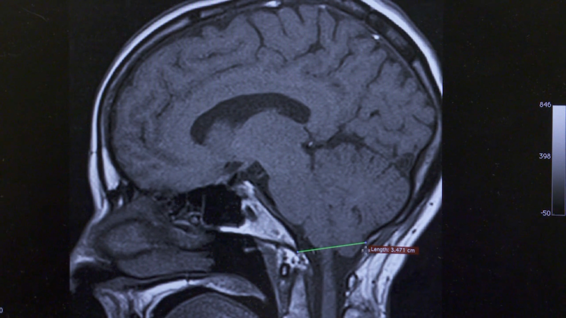
Synopsis. Chiari Malformation Type I (CMI) is a congenital malformation diagnosed by MRI findings of at least 5 mm of cerebellar ectopy below the foramen magnum. CM1 is frequently associated with syringomyelia. Herein, we discuss the history of CMI and syringomyelia, including early pathologic and surgical studies.